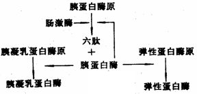
[Principle] In addition to trypsin, which is naturally present in the pancreas of animals, there are two other proteolytic enzymes that share similar properties: chymotrypsin and elastase. During the extraction process, these three enzymes are difficult to separate due to their similar characteristics. Therefore, further purification techniques are necessary to isolate them effectively. Trypsin is typically extracted from the animal’s pancreas. The inactive precursor, trypsinogen, is usually isolated using a dilute acid solution. Then, the pH of the extract is adjusted to around 3.0 using the principle of isoelectric precipitation, causing a large amount of acidic proteins, including trypsinogen, chymotrypsinogen, and elastase, to precipitate out. Ammonium sulfate fractionation is then used to further purify these zymogens. After dissolving the precipitate in water, the pH is raised to approximately 8.0. A small amount of active trypsin is added to activate the zymogens, initiating the conversion of trypsinogen into its active form. Similarly, chymotrypsinogen and elastase are also activated by trypsin in the same solution. [Reagents and Equipment] 1. Reagents (1) Acetic acid solution with a pH of 2.5 to 3.0 (2) 10% acetic acid (by volume) (3) 2 mol/L sulfuric acid (4) Solid ammonium sulfate (5) Anhydrous CaCl₂ (6) Crystalline trypsin (7) 25% ethanol solution containing 0.015 mol/L HCl and 0.05 mol/L CaCl₂ (8) 95% ethanol (by volume) (9) 5 mol/L NaOH (10) Acetone 2. Equipment (1) Fresh pancreas (2) Tissue homogenizer (3) Centrifuge (4) Magnetic stirrer (5) Dialysis bag, 20-mesh sieve, gauze, water bath (6) Beakers, measuring cylinders, graduated pipettes, test tubes, glass funnels (7) Buchner funnel, vacuum filtration flask, thermometer, dropper, magnetic stir bar, gauze, pH paper, etc. [Methods and Steps] Method One 1. Extraction of Trypsinogen Begin by taking about 150g of fresh pancreas, removing connective tissue and fat, and weighing 100g of the clean tissue. Cut it into small pieces and mash it using a tissue homogenizer. Add twice the volume of pre-cooled acetic acid solution (pH 2.5–3.0) and homogenize thoroughly. Transfer the mixture into a 500 mL beaker and adjust the pH to between 2.5 and 3.0 using 10% acetic acid. Extract the mixture at 5–10°C for more than 6 hours, stirring intermittently. Filter the mixture through four layers of gauze and press to collect as much filtrate as possible. Repeat the extraction with an additional 0.5 times the volume of pre-cooled acetic acid solution (about 50 mL), and filter again. Combine the two filtrates and adjust the pH to 2.5–3.0 using 2.5 mol/L sulfuric acid. Let it stand at 4°C for 4 hours, maintaining the pH throughout. Filter the solution using folded filter paper, collect the filtrate, and measure the volume (approximately 200 mL). Add solid ammonium sulfate to the filtrate until it reaches 75% saturation (492 g per liter of filtrate at 5°C). Allow it to stand overnight for complete precipitation of trypsinogen. Next day, perform suction filtration, discard the supernatant, and collect the precipitate as crude trypsinogen. 2. Activation of Trypsinogen Weigh the crude trypsinogen (wet weight) and add 10 times the volume of pre-cooled distilled water to dissolve it completely. Adjust the pH to 8.0 using 5 mol/L NaOH. Add solid anhydrous CaCl₂ while stirring to reach a final Ca²⺠concentration of 0.1 mol/L. Take a 2 mL sample to measure protein content and enzyme activity before activation. Add 5 mg of crystalline trypsin to the solution and gently stir. Incubate at 25°C for 2–4 hours or at 4°C for 12–16 hours. Monitor the increase in trypsin activity every hour until the rate slows down. The specific activity should reach 3,500–4,000 BAEE units/mg. After activation, take another 2 mL sample for analysis. Adjust the pH back to 2.5–3.0 using 2 mol/L sulfuric acid, filter through filter paper, remove the calcium sulfate precipitate, and store the filtrate at 4°C for later use. Method Two Take 100g of frozen pig pancreas and thaw it at 2–5°C. Cut it into small pieces and homogenize using a tissue mincer. Store the mixture at 5–10°C for over 24 hours to allow natural activation of trypsin. Add 250 mL of 25% ethanol solution (containing 0.015 mol/L HCl and 0.05 mol/L CaCl₂) and homogenize for 1 minute. Pour the mixture into a 500 mL beaker, stir intermittently at around 20°C for 5–6 hours. At 1.5 hours, take 2 mL of the activation solution to measure protein content and enzyme activity after activation. After the reaction is complete, centrifuge the mixture at 3,500 rpm for 20 minutes. Filter the supernatant through two layers of gauze. Measure the volume of the filtrate and add powdered ammonium sulfate to bring the solution to 20% saturation. Let it stand for 6 hours, then centrifuge again at 3,500 rpm for 20 minutes. Collect the supernatant for further use and wash the precipitate with 95% ethanol and acetone. Dry the precipitate on a watch glass to obtain crude trypsin I. In the remaining supernatant, add powdered ammonium sulfate to achieve 55% saturation. Let it stand for 6 hours, then centrifuge at 3,500 rpm for 30 minutes. Wash the precipitate with 95% ethanol and acetone, and dry it on a watch glass to obtain crude trypsin II. Foshan Gruwill Hardware Products Co., Ltd. , https://www.zsgruwill.com Another method involves extracting trypsinogen directly from the pancreas. By adjusting the pH of the extraction medium, autolysis of the trypsinogen can be encouraged. In the presence of calcium ions, this process helps the trypsinogen to self-digest slightly and become active under the influence of a small amount of trypsin. This activation step is crucial for obtaining the active enzyme. Once activated, the enzyme solution undergoes further separation and purification steps to isolate trypsin from the other proteolytic enzymes.